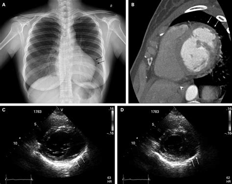
lv diverticulum|lv apical diverticulum : 2024-09-23 Nov 23, 2012 — All diverticula were located in the LV and were most common along the inferior or inferoseptal wall, primarily at the right ventricular insertion . Lage sneakers Merk: Adidas Kleur: Zwart Maat: 40. Lage sneakers Merk: Adidas Kleur: Zwart Maat: 40 Ons verhaal Vacatures Contact Blog Bristol Family. Onze winkels. Nieuw. Dames .
0 · myocardial cleft echo
1 · lv diverticulum vs aneurysm
2 · lv apical diverticulum
3 · lv aneurysm on echo
4 · left ventricular diverticulum echo
5 · left ventricular diverticulum
6 · left ventricular apical diverticulum
7 · congenital diverticulum of left ventricle
8 · More
1 apr. 2022 — How much does it cost to service your Audemars Piguet? It’s difficult to give a cost because a lot depends on the style and model, the age and the condition. But for an .
lv diverticulum*******Jun 1, 2022 — Congenital left ventricular diverticulum (LVD) is a rare congenital anomaly with a prevalence of 0.4% or 3 in 750 cardiac death autopsies , , .A left ventricular diverticulum is .
Nov 23, 2012 — All diverticula were located in the LV and were most common along the inferior or inferoseptal wall, primarily at the right ventricular insertion .

, The dimensions of the described diverticula or aneurysms ranges from 0.5 cm of diameter up to a size of 8 x 9 cm., Most frequently, location of LVA is the LV-apex (28%) and the perivalvular .
Sep 17, 2021 — On the other hand, nonapical diverticula are generally described as isolated cardiac defects of multiple shapes and sizes (ranging from 0.5 cm to 9 cm), with a narrow or .Abstract. Congenital left ventricular (LV) diverticula are rare findings, particularly when first diagnosed in adulthood. We describe successful surgical repair of an isolated congenital .

Jul 1, 2010 — Approximately 30% of LV diverticulum cases are not associated with other congenital malformations, being so-called isolated ventricular diverticulum. Usually, LV .Jul 3, 2013 — Left ventricular diverticulum is an extremely rare anomaly, especially in the absence of other findings, and as such it has been rarely imaged, rarely seen intraoperatively, and has .Mar 5, 2023 — Treatments include resection of the diverticulum, due to risk of spontaneous rupture and systemic embolization, and symptomatic control with beta-blockade, magnesium, ., The dimensions of the described diverticula or aneurysms ranges from 0.5 cm of diameter up to a size of 8 x 9 cm., Most frequently, location of LVA is the LV-apex (28%) and the perivalvular area (close to the mitral valve [= sub-mitral]; 49%) and LVD are found at the LV-apex (57%).lv diverticulumJan 13, 2022 — Introduction. Congenital left ventricle (LV) diverticulum (LVD) is a rare cardiac malformation with a prevalence of approximately 0.1% of all congenital heart diseases. 1 It is characterized by replacement of normal myocardial tissue with either fibrous tissue that subsequently bulges into an outpouching or muscular tissue that results in the formation of a .
Left ventricular (LV) diverticulum is a congenital anomaly that may be clinically silent or associated with various cardiac complications.1 It was originally described in 1838, at autopsy.1 Today, the diagnosis is usually made by means of contrast ventriculography or echocardiography, whereas magnetic resonance imaging is also useful in evaluating this anomaly.
Congenital left ventricular (LV) diverticula are rare findings, particularly when first diagnosed in adulthood. We describe successful surgical repair of an isolated congenital apical LV diverticulum associated with an abnormal submitral apparatus in a young adult who received his diagnosis following a peripheral embolism.
Congenital subaortic left ventricular diverticulum (LVD) is a rare congenital malformation consisting of a localized outpouching from the free wall of the left ventricular outflow tract. The majority of cardiac diverticula arises from the apex of the left ventricle (LV); however, nonapical LVD also occurs, but infrequently. The diagnosis of a muscular diverticulum should be .lv diverticulum lv apical diverticulumJul 30, 2019 — fibrous diverticulum: composed of mostly fibrous tissue; Associations They can be associated with other anatomic defects that involve the thoracoabdominal midline, and have syndromic associations such as pentalogy of Cantrell. Apical diverticula have a higher association with other anomalies. Diverticula occur in isolation in around 30% of cases.Two-dimensional and Doppler echocardiography are sensitive methods for detecting a diverticulum in the interventricular septum, LV wall, subvalvular area, or congenital LV diverticulum (11-13). The case of our patient shows that the diagnosis of congenital LV diverticulum is facilitated only by transthoracic echocardiography.Sep 8, 2017 — In terms of LV myocardial diseases, these diverticulum may be congenital or acquired. Hence, the differential diagnosis of LV myocardial diseases includes aneurysm, pseudoaneur-ysm, diverticulum and LV noncompaction. LVD is a very rare condition in adult and was first described in the 1800s [7,8,9].May 1, 2017 — At 24 months follow-up, the patient was asymptomatic, with normal LV systolic function and free of ventricular arrhythmias. 3. Discussion. The differential diagnosis of left ventricular outpouchings includes aneurysm, pseudoaneurysm and diverticulum [1], [2], [3].According to the Marijon et al. [3] CVD and aneurysm represent two distinct entities, with .Sep 1, 2021 — Congenital LV diverticulum is a rare defect, accounting for 0.05% to 0,7% of all cardiac malformations, according to the different imaging techniques. It is either isolated from or associated with midline thoraco-abdominal abnormalities as part of Cantrell’s syndrome, .A congenital left ventricular aneurysm or diverticulum is a rare cardiac malformation; 411 cases have been reported since its first description in 1816, and other cardiac, vascular or thoraco-abdominal abnormalities have been shown in about 70%. It appears to be a developmental anomaly, starting in the 4th embryonic week.Apr 18, 2011 — LV diverticulum is present in 20% to 50% of these cases . Apical ventricular remodelling. Cardiac remodelling refers to the change in size, shape and function of the heart after injury to the ventricles [16, 21, 22]. The injury is typically due to large acute myocardial infarction depending on anterior descending coronary artery .Oct 22, 2018 — Congenital LV diverticula are characterised by an outpouching that contains endocardium, myocardium, and pericardium, and they are a narrow connection to the cavity and collapse at end-systole. The primary diagnostic feature to differentiate diverticula from crypts is that the congenital diverticula extend outside the epicardial border while crypts remain .May 18, 2010 — The differential diagnosis for a LV outpouching that contracts synchronously with the rest of the ventricle includes an LV diverticulum or an accessory chamber. A diverticulum contains all 3 layers of cardiac tissue but has a narrow connection to the ventricle.
Aug 1, 2022 — Congenital left ventricular diverticulum (LVD) is a rare congenital anomaly with a prevalence of 0.4% or 3 in 750 cardiac death autopsies [1], [2], [3].A left ventricular diverticulum is defined as an enlarged structure containing the endocardium, myocardium, and pericardium and displays normal systolic contraction [4, 5].The left ventricular diverticulum is often .Dec 17, 2019 — Congenital left ventricular diverticula are rare cardiac malformations. In our case a diverticular apical lesion of the left ventricle was accidentally found on a thoracic CT scan, acutely performed to rule out abdominal aortic rupture. . LV = left ventricle; RV = right ventricle Figure 3 Left ventriculogram: the examination showed .
Nov 11, 2018 — This included diverticulum not extending beyond the free wall of the ventricle, often referred to as clefts, and not considered diverticulum in most classification systems . This increasing prevalence and changing location with advancing age at presentation suggests that diverticulum and aneurysms of the cardiac free wall may develop later in adulthood as a .
lv apical diverticulumFeb 11, 2022 — Diverticula are typically narrow-mouthed with a wide outpouching extending beyond the LV myocardial margin—a feature that helps to differentiate them from clefts as previously noted. 5 LV diverticula following myocardial infarction are rarely reported, 6 and are believed to result from incomplete LV rupture caused by hemorrhagic dissection. A gradual .
adidas Lite Racer Rbn 2.0 Sneakers voor heren, zwart, 40.50 EU. 4,2 25 beoordelingen. | Zoek op deze pagina. Momenteel niet verkrijgbaar. We weten niet of en wanneer dit .
lv diverticulum|lv apical diverticulum


